 Discover the intricacies of honey bee anatomy

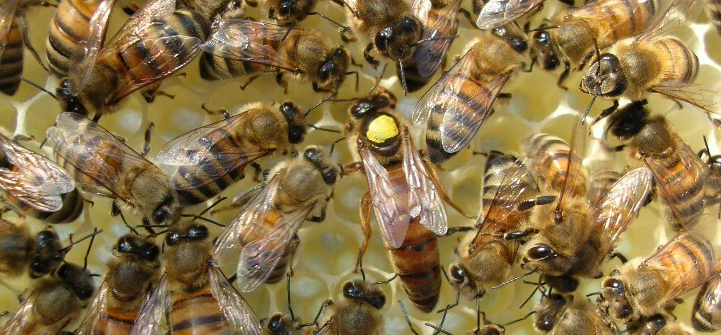
As a member of the insect class (Insecta), honey bees share with other insects the following characteristics. Honey bees are segmented in nearly all their body parts: three segments of thorax, six visible segments of abdomen (the other three are modified into the sting, legs and antenna are also segmented. Honey bees have an exoskeleton, which is rigid and covered with layers of wax, but have no internal bones like vertebrates do. The main component of exoskeleton is chitin which is a polymer of glucose and can support a lot of weight with very little material. The wax layers protect bees from desiccation (losing water). The advantage of chitin-containing exoskeleton also prevents bees from growing continually, instead, they must shed their skins periodically during larval stages, and stay the same size during the adult stage. Bees also have an open circulatory system, meaning that they do not have veins or arteries, but rather all their internal organ are bathed in a liquid called ‘hemolymph’ (a mix of blood and lymphatic fluid). Bees breathe through a complex structure of network of tracheas and air sacs. Oxygen is vacuumed into the body through openings on each segment (spiracles) by the expansion of the air sacs, then the spiracles are closed and air sacs are compressed to force the air into smaller tracheas, which become smaller and smaller until individual tubules reach individual cells. In the following I will discuss the important structures on and inside the honey bee body.
- Table of Contents
- Head Segment of the Honey Bee
- Thorax of the Honey Bee
- Abdomen of the Honey Bee
- Historical Anatomical Literature of Honey Bee Anatomy
Source:
Page text and photos authored and Copyrighted to Zachary Huang, Dept. Entomology, Michigan State University.